

 Dementia Support Line
Research highlight
Using MRI to spot changes in Alzheimer’s disease before symptoms start
Dr Colin Reveley, one of our Daphne Jackson Fellows, is developing MRI techniques to investigate the structure of grey matter in the brain. This could help diagnose dementia sooner and identify those most at risk earlier in life.
Why do we need better MRI of the brain’s grey matter?
My research focuses on grey matter, the crinkled sheet covering the outer surface of the brain. It contains billions of brain cells, and the trillions of connections between them which give us our rich experiences of ourselves, our families, our memories and the world around us. In dementias like Alzheimer’s disease, grey matter breaks down and we can lose those experiences.
In the early stages of dementia, or in the years before symptoms emerge, brain cells in grey matter start to show small changes which can be seen under a microscope. As dementia progresses, the whole grey matter visibly shrinks.
Current hospital MRI scans can show by how much the grey matter has shrunk, but the scanner can’t see objects smaller than about 1 mm. Brain cells are hundreds of times smaller than that, so to understand more about a person’s dementia, both before and after symptoms appear, we need to probe deeper into the grey matter.
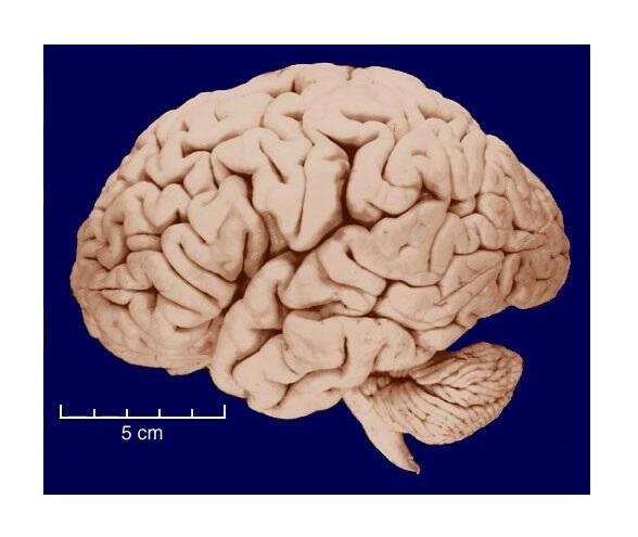
Grey matter is the brain’s crinkled outer surface, imaged adapted from Human (Homo sapiens), University of Wisconsin-Madison Brain Collection
Measuring cell damage before dementia symptoms emerge
At the Oxford Centre for Integrative Neuroimaging (OxCIN), we’re pushing MRI scanning further, to reveal the tiny changes in grey matter brain cells that happen very early in the course of dementia, or potentially even before it starts.
We hope this will help us understand the causes of dementia and assess who is most at risk earlier in life, which could help prevent it.
If the scans work reliably enough, we hope to develop them for those who are already living with dementia, to give more detailed information about its progression, and how well it’s responding to new treatments.
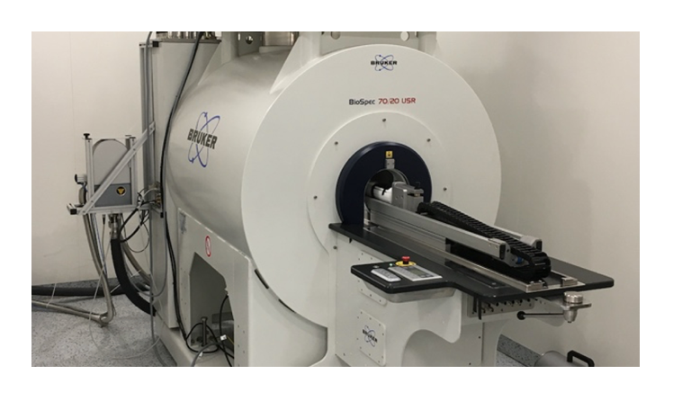
At the Oxford Centre for Integrative Neuroimaging, a specialised MRI scanner will reveal microscopic changes in grey matter. Image credits: John Cairns and Oxford Centre for Integrative Neuroimaging
How the brain scan will work
Although the scanner can’t reveal objects smaller than 1mm, we can use the way water behaves to see deeper into the brain. A glass of water looks still, but at a microscopic level, individual water molecules are frantically moving around at random. In the brain, the water molecules bump into parts of cells and this changes how they move. The scanner can detect these tiny water movements, and we think cells damaged by dementia will affect the water movements differently to healthy cells.
Before we can use this method, I first need to show how the water movements are changed by brain cells. I’ve found that the scans are sensitive to parts of brain cells called 'neurites'. What’s more, they can distinguish neurites that are covered in a fatty substance called 'myelin' from those that aren’t. We know that neurites break down in dementia and that changes in myelin can happen many years before symptoms develop, so the scans could help predict who is most at risk.
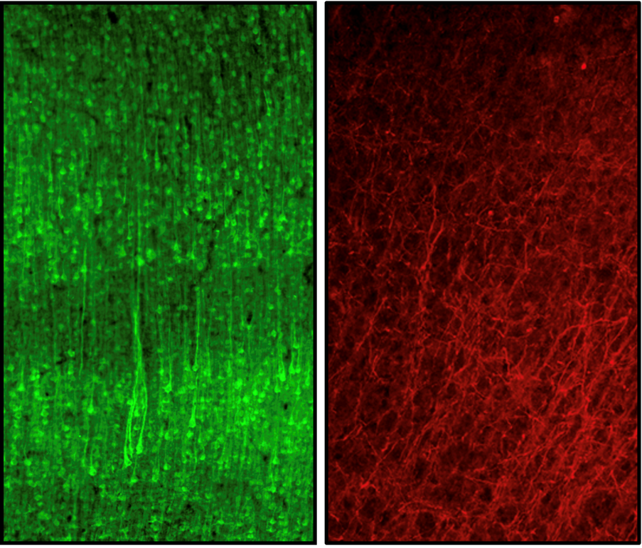
Neurites showing dendrites (left) and axons covered in myelin (right) under a microscope. Image adapted from Reveley et al. (2022). doi: 10.1038/s41467-022-34328-z
People’s brains differ in lots of ways, so even if their scans are different, I can’t be certain why. To confirm that the scans are showing changes in myelin, not something else, I gave mice a treatment to reduce the amount of myelin in their grey matter before scanning them. The treated mice didn’t change their behaviour, and we carefully monitor their welfare. When I compared treated and untreated mice, I was able to confirm the changes in myelin caused the changes in the scans.
Moving towards clinical use
The next step is to check that the same differences we now know are caused by myelin changes are visible when I scan small brain samples donated to the Oxford Brain Bank by people who lived with dementia. In mice, the diffusion patterns may also reveal changes in inflammation, which we know is important in dementia, so I am now investigating that too.
We don’t have to wait to test the scans in people living with dementia. While I am investigating how the scans work using mice and human samples other researchers in OxCIN are developing them for use in hospital scanners and getting ready to test them in people living with dementia.
Daphne Jackson Trust Fellowship
I'm funded by Alzheimer’s Society through a Daphne Jackson Fellowship, which is a unique way of returning to scientific research after a period away due to illness or caring responsibilities.
This fellowship allowed me to lead my own research, and because it’s part time it allowed me to ease gradually back into research. After a year working part time, the University of Oxford agreed to provide extra funding to expand my research.
What research are we supporting?
Alzheimer’s Society is one of the UK’s biggest charitable funders of dementia research – funding over £120 million of dementia research since 1989.